EUR 0,00
(Delivery cost: 0,00)


The function requires that you are logged in. Create a user and use our smart list system to keep track of future purchases, regular purchases or wish lists.
The differential interference contrast (DIC) device is used to study transparent low-contrast specimens that are not visible in the bright field.
The DIC technique introduces a pseudo-relief image effect. The color and intensity effects in the image are determined by the rate of change of the refractive index and the thickness of adjacent sections of the sample. The image of the sample appears three-dimensional. The 3D view does not show the actual geometry of the specimen; it colors the specimen according to the optical thickness. This technique is not suitable for measuring the actual height and depth.
The DIC and phase-contrast techniques complement each other. They produce similar contrast images, but the DIC images have no diffraction phase contrast halos around specimens. These halos result from the difference between the refractive index of the sample and the surrounding medium. They often obscure the details at the edges of the observed structures. The DIC technique allows you to see these details.
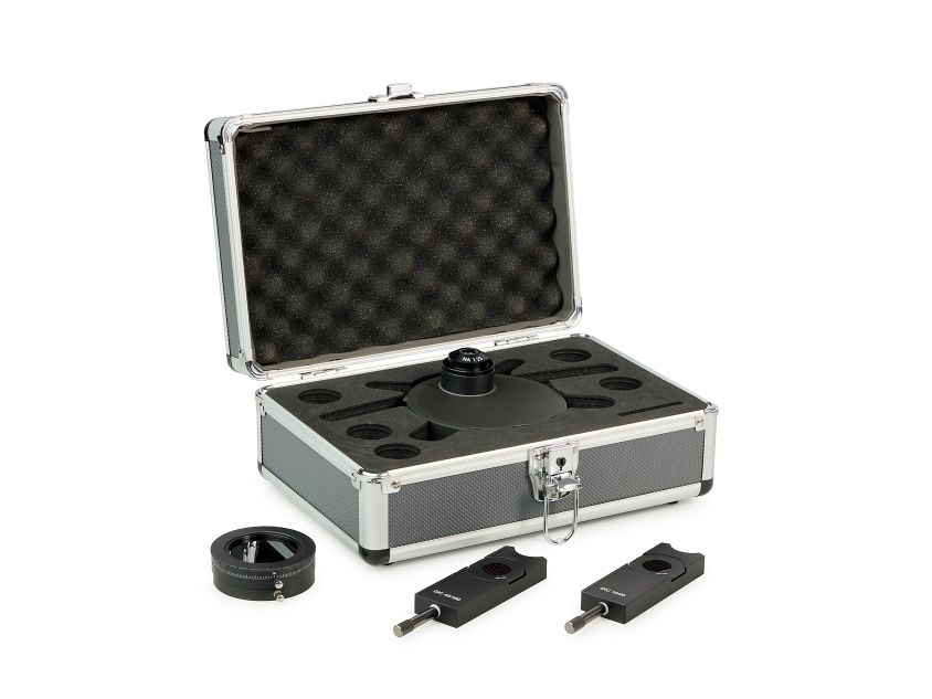
The differential-interference contrast device includes a DIC condenser, polarization filter, and DIC sliders.
DIC condenser
The DIC condenser is installed in place of the brightfield condenser. The turret includes DIC prisms and a slot that is used during brightfield observations. The DIC prisms in the turret are used depending on the objective being used. Under the turret, there is an aperture diaphragm adjustment control, which is used when working in brightfield.
DIC sliders
The DIC slider is installed in a special slot above the revolving nosepiece. The DIC slider is used in combination with the corresponding objective. The slider is a plate with a DIC prism and an analyzer. The MAGUS DIC290 device includes two sliders: One is used with 10x and 20x objectives, and the other one with 40x and 100x objectives. The rotation of the slider knob moves the DIC prism in the transverse direction.
Polarization filter
When the DIC technique is used, the light from the illuminator is passed through a polarization filter. The polarizer is placed on the collector and secured with a knurled-head screw. The polarizer is marked with a rotation angle scale and can be rotated 360°. The intensity of the transmitted light changes depending on how the polarizer and analyzer are positioned relative to each other. When they are mutually perpendicular to each other, a complete darkening effect is observed. The polarizer can be flipped down: You do not have to take it off the microscope to remove it from the optical path.
Recommended objectives
The DIC technique requires a precise position of the optical components relative to each other: the lower DIC prism – relative to the front focal plane of the condenser and the upper DIC prism – relative to the rear focal plane of the objective. For DIC observations, you can use the objectives that are supplied with the microscope or additional MAGUS FL S-APO60 PlanF objectives with a higher aperture.
A higher aperture of an objective allows focusing on a thin flat section of a thick specimen, which is called optical sectioning. Higher apertures also increase the resolving power of the optical microscope.
Compatibility:
Package:
| Brand | MAGUS |
| Warranty | 2 years |
| EAN | 5905555025629 |
| Package size (LxWxH) | 4x7x4 cm |